enes

Blog

A common in-office procedure is an eye injection for the treatment of various eye conditions, such as wet macular degeneration, diabetic retinopathy, or vein occlusion.  

It is normal to be anxious prior to your first eye injection but knowing what to expect may alleviate some anxiety.  

Your eye will be numbed with topical anesthetic drops. Usually, a cotton-tipped applicator soaked in anesthetic will be held against the white part of the eye to numb it.  Sometimes, a thick gel may also be used for numbing. Once the eye is numbed, it will then be cleaned and a small instrument will be used to hold the eye open. During the actual injection, some patients feel pressure while others may feel a quick sting. Each person will have a different experience and each injection will be different.

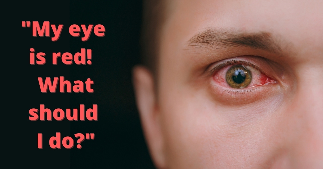
The medicine is injected into the white part of the eye. Sometimes a blood vessel may be encountered during the injection and you may notice a little redness on the outside of the eye. This is purely a cosmetic symptom and the redness will disappear within the next couple of days. Patients may also experience some mild irritation after the injection due to the drops that are placed in the eye. I usually recommend over-the-counter artificial tears to use after the injections. Usually, the eye should feel normal by the next day.  

Another common symptom after an injection is floaters. These floaters usually appear as small bubbles in the lower half of your vision, but they will disappear in a day or two.  

There are no activity restrictions after the injection and no antibiotic drops are needed.  

Things to watch for after the injection:

- If your eye gets painful or redness develops a couple of days after the injection, then you should call your doctor.  

- If your vision declines or you notice increased or hundreds of floaters then you should call to be seen.

Otherwise, patients are usually initially seen once a month after they first start injections

Article contributed by Dr. Jane Pan

This blog provides general information and discussion about eye health and related subjects. The words and other content provided in this blog, and in any linked materials, are not intended and should not be construed as medical advice. If the reader or any other person has a medical concern, he or she should consult with an appropriately licensed physician. The content of this blog cannot be reproduced or duplicated without the express written consent of Eye IQ.

Diabetic retinopathy, which is a complication of diabetes that affects the eyes, is detected during a comprehensive eye exam that includes:

  • Visual acuity testing.
  • Dilated exam in which drops are placed to widen the pupil to allow examination of the retina.
  • Tonometry. Measurement of the eye pressure inside the eye.

Supplemental testing may include:

  • Optical coherence tomography (OCT). This is a non-invasive test that images the retina to detect any fluid or diabetic macular edema.
  • Fluorescein Angiography. This test involves an injection of a dye into your arm and a series of pictures that are taken as the dye flows through the retinal vessels. This may show leakage of fluid or the growth of new blood vessels in the retina.

Treatment for Diabetic Retinopathy

The best treatment is prevention of diabetic retinopathy by strict control of blood sugars. Once diabetic retinopathy is present, treatment of diabetic retinopathy will slow progression but won’t cure it.

Diabetic macular edema can be treated with several different therapies that may be used alone or in combination.

These include:

  • Injection therapy. Anti-VEGF drugs are injected into the eye to block a substance called vascular endothelial growth factor (VEGF). This medication will block abnormal blood vessel growth and decrease the leakage of fluid into the retina. The three most commonly used drugs include Avastin, Lucentis and Eylea. Steroids are another option for treatment of diabetic macular edema but are used less often due to side effects of possible cataracts and increased eye pressure.
  • Laser therapy. Small laser burns are applied in the area of the retinal swelling to slow the leakage of fluid. Laser treatment can be combined with anti-VEGF injections.

Proliferative diabetic retinopathy can be treated with:

  • Laser therapy. Laser is applied on the retina to shrink the new blood vessels and to prevent bleeding inside the eye. Bleeding inside the eye will cloud the vision and cause floaters, which are small moving spots that appear in your field of vision.
  • Surgery. When there is a significant amount of blood or scar tissue inside the eye, then vitrectomy surgery is performed to remove the blood and scar tissue. Laser and anti-VEGF therapy may also be applied during the surgery.

In the end, the best treatment is prevention of diabetic retinopathy. An annual comprehensive dilated eye exam is recommended for anyone who has been diagnosed with diabetes, since early mild non-proliferative diabetic retinopathy can show no symptoms.


Article contributed by Jane Pan M.D.

The content of this blog cannot be reproduced or duplicated without the express written consent of Eye IQ

bcm_videoplayer_banner

Video Education Library

Click the button below to view our entire collection of patient educational videos. Our video library covers a range of eye related topics including vision conditions, optical procedures, routine eye exams.

Watch Videos